|
|||
AbstractMaloccluded teeth need orthodontic treatment. But in some cases it seems difficult to justify the need for orthodontic treatment in the dental clinics and hospitals with out using the diagnostic tools especially at the remote areas. This ultimately increases the number of orthodontic patients seeking treatment and depriving the genuine patients from receiving treatment in proper time. Proper diagnosis and early intervention of malocclusion in dental clinics and hospitals, with very minimum efforts could be a key to prevent this unwanted situation. This article therefore was designed to discuss these practical oriented and frequently encountered problems in the dental clinics and hospitals of Bangladesh and to find out a guide line to an easy solution of it.
|
|||
| (Pioneer Dental Journal, Vol.02, No.01.) | |||
IntroductionNormal occlusion and malocclusion are two opposite side of a coin. However, there are some cases where it is difficult to differentiate normal occlusion from malocclusion. Normally it is very easy to notice malocclusion in patients but in some cases other features make it difficult to identify as malocclusion. Before we discuss the present article it would be logical to focus a little on different aspect of normal occlusion and malocclusion for easy understanding of the readers.
|
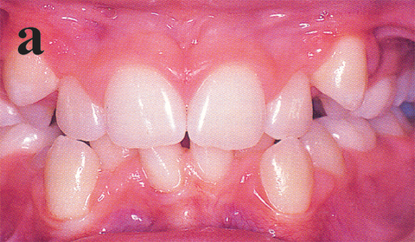
What is Normal Occlusion?It is well known among the dentists that Angle’s Class I molar relationship along with class I canine and incisor relationship with 2-3 mm overjet and overbite should be considered as normal or ideal occlusion. But there are some patients who have all class I dental features but some minor irregularities push them into malocclusion group. These types of cases sometimes put the dentists in dilemma to decide whether the patients need treatment or not. What is Malocclusion? An unacceptable deviation- esthetically and/or functionally from the ideal occlusion is called Malocclusion. (Fig:1a). However, the term “Unacceptable deviation” sometimes used to make the dentists confused to differentiate the malocclusion from that of normal occlusion as there was no such easy method to diagnose this type of case with out the diagnostic tools like model casts, orthopentamogram and lateral cephalogram. But in practice sometimes it becomes very important to justify the case on the spot only by the visual appearance of the patient1. Therefore, it was inevitable that an easy method should be available in practice to find out who really needs orthodontic treatment. |
||
| PDJ, Vol-02, No-01, January 2018 | |||
|
|||
|
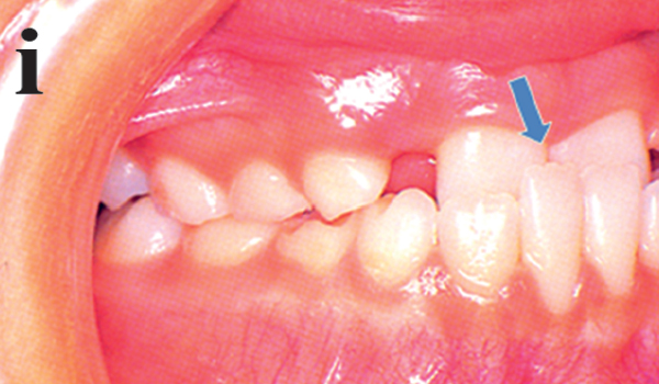
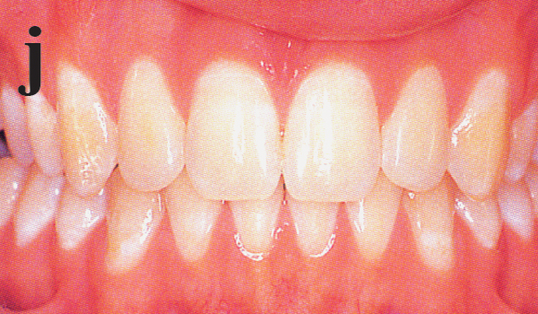
Problems those lead the patients to seek for the orthodontic treatment: Protruded, irregular or maloccluded teeth can cause three types of problems: 1. Psychosocial problems: Patients suffer from inferiority complex. Patients become socially handicap. They face negative status in the schools and in the employability. They even face negative status during competition for a mate. And these problems are not “just cosmetic”. 2. Oral functions: A severe malocclusion may compromise all aspects of oral functions. Adults with severe malocclusion routinely report difficulty in chewing, which after treatment are largely corrected. It can be difficult or impossible to produce certain sounds and effective speech therapy may need some orthodontic treatment. Relationship of malocclusion and adaptive function to Temporo-mandibular dysfunctions (TMDs) manifested as pain in and around the TM joint take place in many patients.1,33 3. Relationship to injury and dental diseases: Malocclusion has a deep relation to different dental injuries and diseases. Protruded maxillary incisors can increase the likelihood of injury in class II cases resulting in fracture or devitalisation of pulp. Extreme overbite leads the lower incisors to contact the palate and can cause significant tissue damages and ultimately loss of upper incisors in few patients. Extreme wear of incisors also occur in excessive overbite cases. Malocclusion can contribute to both dental decay and periodontal disease by making harder to care for the teeth properly or by causing occlusal trauma. How to diagnose the treatment need? Severity of a malocclusion correlates with need for treatment. Several indices for scoring the deviation of teeth from the normal as indicator of orthodontic treatment need were proposed. Among them scoring system by Shaw and co-workers the “Index of Treatment Need” (IOTN) seems effective. IOTN places patients in five grades from “No need for Treatment” to “Treatment Need”. The index has a dental health component derived from occlusion and alignment and an esthetic component derived from comparison of dental appearance to some standard photographs. This can help the dentists to find out whether the patients need orthodontic treatment. |
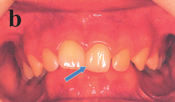
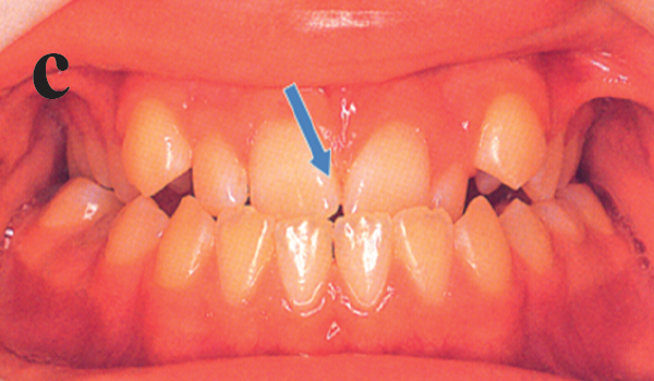
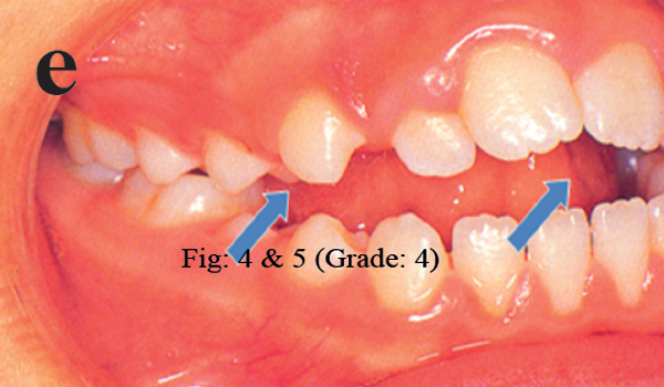
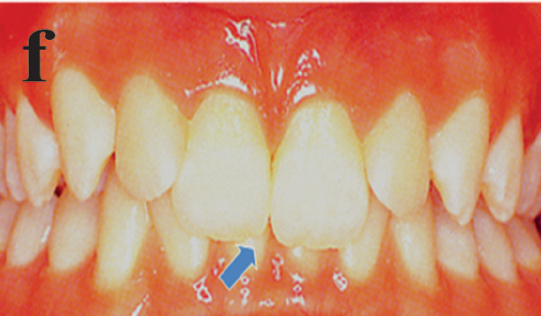
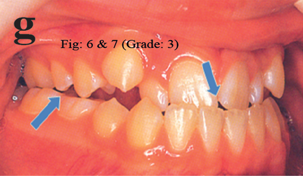
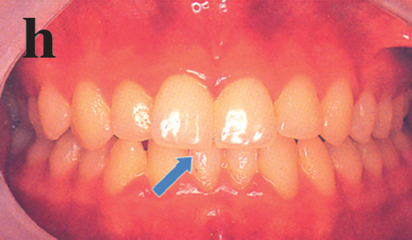
These are as follows: IOTN: Index Of Treatment Need Grade 5 (Extreme/ Need Treatment) Fig:1b, 1c
Extremely minor malocclusions including contact point displacements less than 1 mm. Conclusion: In the present article an effort was made for easy and handy “On the spot diagnosis” of the patients who need orthodontic treatment on the basis of some series of photographs. It might be expected that this widely used method would be proved useful for the dentists in the dental clinics and hospitals in remote areas of our country where the diagnostic tools are not readily available for early diagnosis and proper intervention. Therefore, proper utilization of this easy method would help a great number of orthodontic patients to be diagnosed early to receive the proper treatment thereafter. |
||
| PDJ, Vol-02, No-01, January 2018 | |||
|
|||
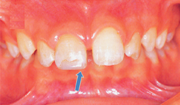
| Figure: a) Malocclusion, b & c (Grade: 5), d & e (Grade: 4) , f & g (Grade: 3), h & i (Grade: 2), j (Grade:1) | |||
References: | |||
|---|---|---|---|
|
|
||
| PDJ, Vol-02, No-01, January 2018 | |||
|
|||
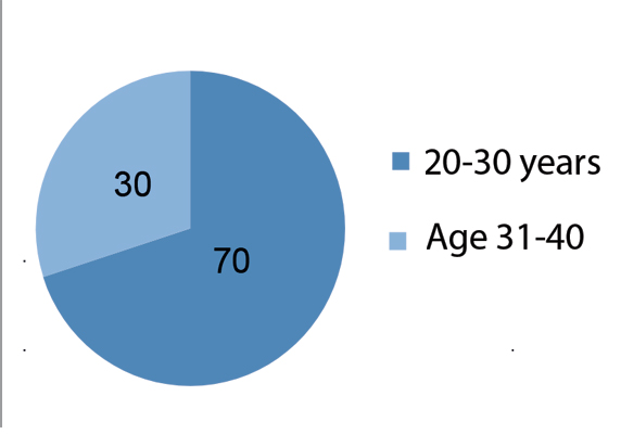
AbstractBackgrounds: For aesthetic and functional outcomes, it is necessary to place the maxillary central incisors in complete denture in the same position as natural teeth, relative to incisive papilla. Methods:61 Patients (male and female) of 20 -40 years age range were selected with dentate maxillae and intact maxillary dental arch. In order to determine what type an individual belongs, we imagine two lines one on either side of the face, running about 2.5 cm in front of the tragus of the ear and through the angle of the jaw. if they diverge at the chin the type is ovoid; if they converge towards the chin the type is tapering. Results: Mean value of central incisor to incisive papilla distance for tapered face formed male was 12.59 mm and 12.35 mm for female, 11.25 mm for ovoid face-formed male and female. Conclusions: Central incisor to incisive papilla distance differs significantly in tapered and ovoid face formed patients.
|
|||
| (Pioneer Dental Journal, Vol.02, No.01.) | |||
IntroductionAfter loss of natural teeth, provision of prosthodontic services almost becomes a necessity in the present day living. Prosthodontists who treat a large number of edentulous patients realize that there are a number of patients who cannot be satisfied aesthetically, functionally or both. For these patients, even a more objective selection criteria will be unsuccessful. However, for the majority of edentulous patients, a simple objective technique involving anatomical measurements would provide at least a starting point for tooth selection. This is more valuable for patients who request denture fabrication and have no previous denture or dental records to utilize for this process.
|
What is Normal Occlusion?Prosthesis cannot be an exact substitute of natural teeth, if prepared properly based upon some measurable parameters then they are not only functionally stable but also aesthetically and biologically viable.12 To improve aesthetic and functional outcomes with respect to patients’ requirements and desires, it is necessary to place the maxillary central incisors in complete denture in the same position as natural teeth, relative to incisive papilla.1 The earlier researchers have described different arch form as square, tapered and ovoid.16 Combinations of these forms are well recognized in prosthodontics.5 The assessment of arch forms has been done by their geometric description.7, 8, 13-15,16 There is a distinct relationship between face form and arch form, especially as related to the upper arch.6 In absence of pre-extraction records, the incisive papilla is an important anatomical landmark that can be used as an aid for anterior teeth positioning.1 It is an immobile structure and usually does not shift in adult life.2,12 The researcher used maxillary central incisor to incisive papilla distance as a biometric guide.3,4,12 With gross resorption of the buccal plate after tooth extraction, the papilla may appear to be on the crest of the alveolar ridge and in cases of more severe resorption it would appear to be in front of the ridge. |
||
| PDJ, Vol-02, No-01, January 2018 | |||
|
|||
|
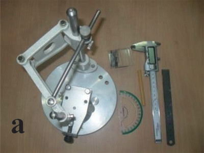
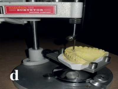
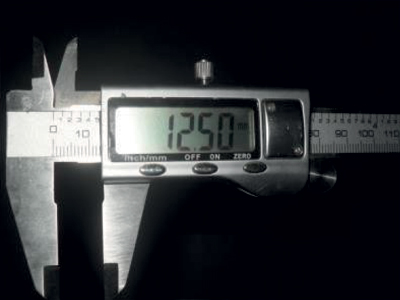
The midpoint of incisive papilla is more commonly used as the reference point, although the posterior part of the incisive papilla is more stable, as it undergoes least change after teeth have been extracted.1,2 This investigation was done to determine whether measurement of central incisor to incisive papilla distance in dentate individuals can provide some meaningful guidelines for the maxillary anterior teeth arrangement while dealing with prosthodontic patients having similar face forms. ObjectiveTo find out the distance between incisive papilla and maxillary central incisors in a tapered face form and a ovoid face form as a guide to anterior teeth arrangement for complete denture. Materials and methodsThe study was designed as cross sectional and observational. The study was done in department of prosthodontics, Bangabandhu Sheikh Mujib Medical University (BSMMU) From March’ 2014 to August’ 2014. Patients attended in the department of prosthodontics in BSMMU for the treatment of the lower arch. It was a non-probability convenience sampling method. 61 Patients (male and female) of 20 -40 years age range were selected with dentate maxillae. Intact maxillary dental arch., periodontally sound maxillary anterior teeth, class I arch relationship, angle’s class I occlusal relationship were in inclusion criteria. Patients with supernumerary tooth erupted in maxillary arch, maxillary midline diastema, any degree of crowding in maxillary dentition, visible attrition involving incisal edge, rotation of maxillary anterior teeth, history of previous orthodontic treatment, any soft tissue lesion involving incisive papilla, no history of surgical procedure in maxillary arch were in exclusion criteria. Digital verniaer scale, calibrated transparent protector, surveyor-Marathon-103, base former, lead pencil, rubber bowl, spatula and impression tray were the equipments. Alginate, die stone and sticky wax were used as materials. Data are expressed as mean, ± standard deviation. Statistical analysis was done by ANOVA. P < 0.05 level of significance was selected for all analyses. ProceduresThe patient was selected by thorough medical and dental history followed by selection criteria of this study. |
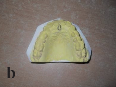
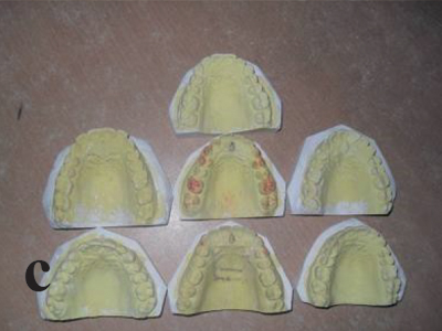
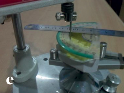
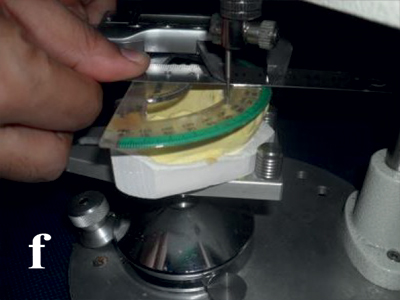
The patient was seated on the well equipped dental chair in free head position. Face form was assessed. Human face form was classified into 3 types: square, tapering and ovoid. Here I selected the face form of ovoid and tapering. In order to determine what type an individual belongs, we imagine two lines one on either side of the face, running about 2.5 cm in front of the tragus of the ear and through the angle of the jaw. if they diverge at the chin the type is ovoid; if they converge towards the chin the type is tapering. After that the impression was taken with high viscosity alginate impression material by using correct powder/liquid ratios, and proper mixing technique as provided by the manufacturer. After taking the impression, it was rinsed in running water. Sterilization of the impression was done. The cast was poured with die stone within 15 minutes after making the impression. Standardization was done by making the base of the cast by using base former. The cast was retrieved from the base former after 45 minutes. Arch form was assessed on the cast. The central incisors in the ovoid arch are forward of the canines in a position between that of square and tapering arch, have a broader effect that should harmonize with an ovoid face. The central incisors in the tapering arch are a greater distance forward from the canines than any other arch. This is usually in harmony with a tapering face. Incisive papilla was first identified and then the boundaries were marked by using a hard lead pencil. Cast was secured to a cast surveyor. The horizontal distance between vertical pin of the surveyor and the embrasure between the maxillary central incisors was measured by placing the protector in such a way that it’s 90 degree marking was almost superimposing the vertical pin of the surveyor, which at this stage was touching the posterior border of incisive papilla. After securing protector in that manner sticky wax was applied to protector and the vertical pin, to stop any unwanted movement. Horizontal distance will be measured on the calibrated transparent protector by using caliper device placing at one end which will coincide with the vertical pin and the other end of the incisal edges. ResultsTotal 61 males and females with 20-40 years old patients were observed for the result of taper and ovoid face form |
||
| PDJ, Vol-02, No-01, January 2018 | |||
|
|||
| |||
| PDJ, Vol-02, No-01, January 2018 | |||
| |||
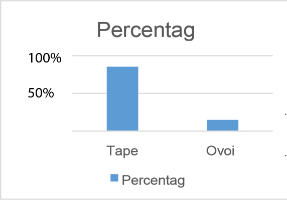
| In figure-2 showed that, 85.25% in taper group and 14.75% in ovoid group. | |||
| PDJ, Vol-02, No-01, January 2018 | |||

